High Resolution Ultrasound

The Center for Fetal Medicine is a state-approved Prenatal Diagnosis Center. We have the ability to screen and test for maternal or fetal complications using state-of-the-art ultrasound (including 3D and 4D capabilities) in conjunction with diagnostic testing as needed. We provide preconception consultations, gynecologic ultrasound evaluations, and advanced care for complicated maternal-fetal conditions and multiple gestations.
Obstetrical ultrasound is a safe and noninvasive imaging method that uses sound waves to create real-time pictures of the fetus, placenta, and amniotic fluid. Ultrasound is typically performed at several points during pregnancy to assess fetal growth and development. In the first trimester, it can confirm gestational age and detect certain early abnormalities. As the pregnancy progresses, ultrasound helps monitor fetal size and position, as well as the health of the placenta and amniotic fluid. This ongoing evaluation supports planning for a safe pregnancy and delivery.
An obstetrical ultrasound may be performed during a routine visit in your doctor’s office. Depending on the stage of pregnancy and the clinical need, the exam may be performed transabdominally or transvaginally. A transabdominal ultrasound involves applying gel to the lower abdomen and moving a handheld transducer over the skin to obtain images. For this type of exam, a full bladder is often required to improve visualization and should not be emptied until after the procedure. A typical transabdominal ultrasound takes approximately 30 to 60 minutes.
A transvaginal ultrasound involves inserting a transducer into the vagina for a comprehensive view of the fetus and surrounding organs. Patients may experience some mild pressure similar to that experienced during a regular gynecological exam. There are no special preparations needed for this procedure, and it can usually be completed in 15 to 30 minutes. A transvaginal ultrasound can provide more detailed images of the uterus and ovaries and is especially useful during the early stages of pregnancy.
Ultrasound images are displayed in real time, allowing both the patient and physician to view the scan as it occurs. Additional detailed interpretation is typically completed within one to two days and reviewed by your doctor. Normal results indicate appropriate fetal growth, normal heart and breathing activity, and no visible abnormalities.
If any concerns are identified, your doctor may recommend further testing or follow-up procedures to better evaluate the findings.
What Technology do we use?
 The Center for Fetal Medicine and Women’s Ultrasound is proud to offer advanced diagnostic imaging using state-of-the-art ultrasound technology. Our facility is fully accredited by the American Institute of Ultrasound in Medicine (AIUM), demonstrating our commitment to the highest standards of quality and safety.
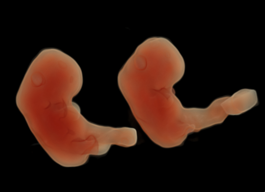
Our team includes board-certified maternal-fetal medicine specialists and highly trained perinatal sonographers. Using sophisticated imaging techniques—including enhanced illumination technology capable of rendering lifelike fetal images with up to three virtual light sources—we are able to provide exceptional detail and clarity.
Our ultrasound services include color and pulsed Doppler studies, fetal biophysical profiles, fetal echocardiography, and nuchal translucency screening, allowing for comprehensive evaluation throughout pregnancy.
High-resolution ultrasound allows for a detailed evaluation of fetal anatomy and can help identify potential structural findings associated with certain genetic or congenital conditions. This level of imaging can assist in assessing the baby’s risk for conditions such as:
-
Spina bifida
-
Cleft lip and palate
-
Congenital heart abnormalities
-
Down syndrome
-
Trisomy 18 and Trisomy 13
-
Skeletal dysplasias
High-resolution ultrasound is a safe, noninvasive imaging technique that uses high-frequency sound waves—not radiation—to evaluate the developing pregnancy. It poses no known risk to either the mother or the baby.